
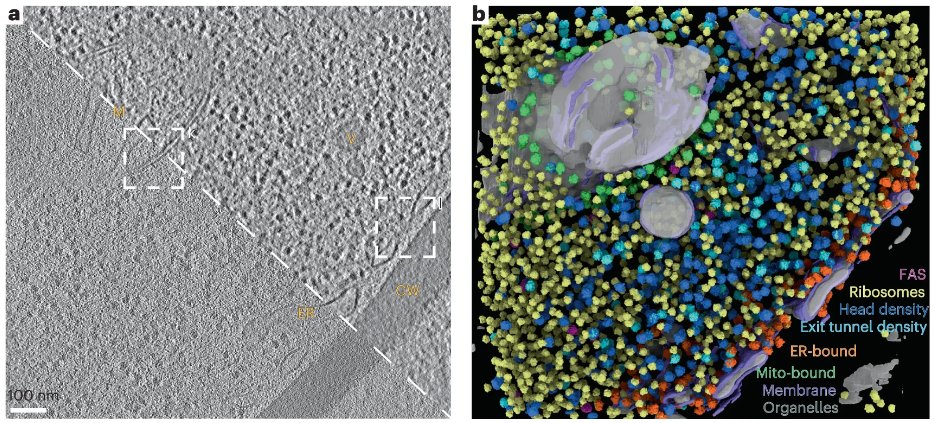
AI Organelle Segmentation is rapidly transforming how scientists analyze microscopic cellular structures. Researchers are using artificial intelligence to improve the accuracy of identifying organelles in live-cell imaging, a critical step for understanding how cells function and respond to biological processes.
AI Organelle Segmentation allows scientists to isolate specific cellular components in microscopy images by distinguishing organelles from surrounding structures, noise, and background signals. This segmentation process produces masks that enable precise quantitative analysis, including measurements of organelle size, shape, movement, and spatial relationships within cells.
As advanced microscopy techniques generate increasingly complex datasets, artificial intelligence has become essential for handling the scale and complexity of biological imaging.
AI Organelle Segmentation supports modern microscopy analysis
In cellular imaging research, AI Organelle Segmentation plays a vital role in interpreting microscopic images. Accurate segmentation enables researchers to track biological events, analyze organelle interactions, and study structural changes inside living cells.
Super-resolution imaging techniques have improved the ability to observe cellular details. However, they also introduce challenges such as imaging noise, signal variation, and phototoxicity effects that can distort biological signals.
Because of these limitations, scientists need segmentation algorithms capable of performing reliably across different imaging platforms and experimental conditions. Artificial intelligence models are increasingly being used to overcome these challenges and deliver consistent results.
AI Organelle Segmentation evolves with deep learning models
Researchers studying AI Organelle Segmentation note that the field has evolved from traditional image-processing techniques to advanced deep learning models.
Earlier segmentation approaches relied on classical image analysis methods. These techniques still remain useful for images with high contrast and clearly defined structures. They are also valuable for rapid screening and generating preliminary labels used in AI training.
However, modern deep learning models now dominate complex segmentation tasks. Architectures such as Fully Convolutional Networks, U-Net, and Mask R-CNN can learn hierarchical patterns within microscopy images.
These models analyze images in an end-to-end process that improves accuracy and robustness. As a result, they can detect complex organelle structures, including filamentous networks and overlapping cellular components.
AI Organelle Segmentation adapts to organelle complexity
A key challenge in AI Organelle Segmentation arises from the diversity of organelle structures. Different cellular components have unique shapes, behaviors, and dynamic patterns.
For example, mitochondria frequently change form as they undergo processes such as fission and fusion. These transitions create networks or punctate structures that require specialized segmentation and tracking algorithms.
Similarly, the endoplasmic reticulum forms complex tubular and sheet-like structures inside cells. Accurately analyzing this network requires segmentation methods that preserve continuity and allow further structural analysis.
Other organelles, including lysosomes, Golgi bodies, and lipid droplets, also vary significantly in size and distribution. Effective segmentation algorithms must therefore adapt to these variations while maintaining accuracy across different biological conditions.
AI Organelle Segmentation enables multi-organelle analysis
Another important development in AI Organelle Segmentation is the transition from analyzing single organelles to studying multiple organelles simultaneously.
Traditional approaches often focused on one organelle type at a time. However, researchers now aim to understand how organelles interact within cellular networks.
This shift requires systems that can segment multiple organelles within the same image while maintaining spatial relationships between them. By doing so, scientists can study how different organelles coordinate biological functions.
Such multi-organelle analysis provides a more comprehensive understanding of intracellular regulation and cellular health.
AI Organelle Segmentation faces key research challenges
Despite major progress, several challenges still limit the widespread adoption of AI Organelle Segmentation.
One issue involves the computational demands of analyzing three-dimensional imaging data. Advanced microscopy often produces large datasets that require powerful computing resources for segmentation.
Another challenge involves the need for extensive labeled training data. Many deep learning models rely heavily on manually annotated datasets, which require significant time and expertise to produce.
Researchers are addressing these issues through several strategies. Self-supervised learning methods can reduce the need for large labeled datasets. Transfer learning allows models trained on one dataset to adapt to new imaging conditions.
In addition, scientists are using synthetic data and physics-based models to improve algorithm robustness. Active learning techniques also help researchers prioritize which samples should be labeled to improve training efficiency.
AI Organelle Segmentation reshapes cell biology research
As artificial intelligence continues to evolve,Organelle Segmentation is becoming more than just a technical tool. It is developing into a scalable research infrastructure that supports advanced biological discovery.
These technologies allow researchers to move beyond qualitative observations toward precise quantitative analysis of cellular processes. By analyzing organelle dynamics at scale, scientists can gain deeper insights into disease mechanisms, cellular behavior, and biological regulation.
In the coming years, AI Organelle Segmentation is expected to become an essential component of modern biomedical research, enabling more accurate and comprehensive exploration of life at the cellular level.


